Mysteries of Cell Fate Unlocked with New Measurement and Modeling Techniques
 isbscience.org/news/2020/04/23/cell-fate/
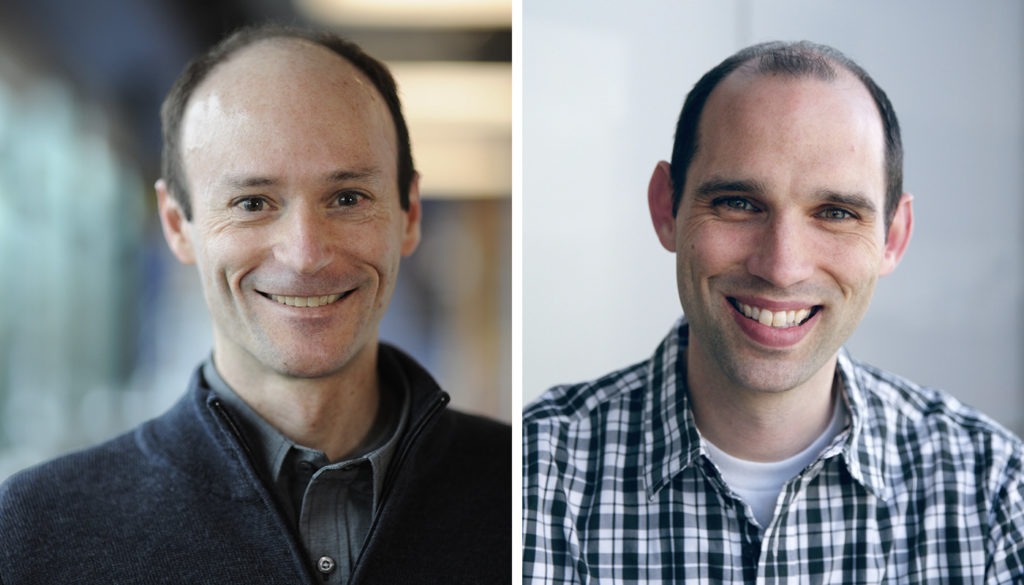
ISB Professor Dr. Jeff Ranish, left, and Research Scientist Dr. Mark Gillespie.
What makes a red blood cell a red blood cell?
The cellular process of differentiation — when a cell becomes specialized — happens inside every living thing. Researchers have known about and studied this process for many years, but information about the concentrations of an important class of proteins residing in a cell’s nucleus has been lacking — a missing link needed to fully understand how the process works.
ISB researchers and collaborators have quantified this important class of nucleus-residing proteins that play a key role in the formation of red blood cells. These proteins, called transcription factors (TFs), turn genes on or off by binding to DNA.
This work is broadly applicable to all cell types, not just red blood cells, as TFs are expressed everywhere and are involved in specifying differentiation.
Scientists have measured TF proteins in prior years, but rarely at the absolute level, or in the systematic way this research team performed. ISB’s team made multiple absolute measurements along a cell’s lineage — from stem cell to red blood cell — providing them insights never seen before.
In research published in the journal Molecular Cell, scientists determined the absolute concentration of 103 TFs and co-factors during the course of red blood cell formation, providing a dynamic and quantitative scale for TFs in the nucleus.
Understanding Cell Fate
The fate of a cell is determined, in part, by the relationship of different TF concentrations within the cell, a concept called stoichiometry.
“We have directly measured TF concentrations and compared them to determine stoichiometry in order to better understand how cells make and execute lineage decisions,” said Dr. Mark Gillespie, co-lead author of the paper and research scientist in ISB’s Ranish Lab.
To quantify these proteins, researchers needed a reliable technology that was sensitive enough to systematically measure low abundance TFs and cofactors from progenitor and precursor cells during human red blood cell differentiation, a.k.a., erythropoiesis. For this, they used a targeted mass spectrometry approach called selected reaction monitoring, or SRM. They adapted the technology such that their measurements would yield absolute quantities of proteins, which is needed to compare the concentrations of different proteins with one another.
Gillespie and his colleagues also established the first gene regulatory network of cell fate commitment that combines temporal protein concentration data with mRNA measurements. The researchers were surprised to discover that, in the nucleus, co-repressors are dramatically more abundant than co-activators at the protein, but not at the RNA level, a finding that is likely a general principle underlying cell biology
“The dynamic modeling and TF concentration measurements this team created provide valuable resources for researchers around the world to explore biological territory we simply could not before,” said ISB Professor Dr. Jeff Ranish, one of the corresponding authors of the paper. “We plan to build upon the results of this exciting study to enable comprehensive measurements of the TF proteome during differentiation and to be able to provide TF concentrations in single cells.
This work was a collaboration between ISB, Ottawa Hospital Research Institute, University of Ottawa, University of Bern, University of Oxford, and University of Washington.


