Feb. 27, 2017
3 Bullets:
- A multi-institute collaboration mapped in high resolution the earliest effects of the Huntington’s disease mutation in mice
- The study included four different genetic strains of mice which allowed the researchers to observe differences in the rate of mutation-induced changes as a result of genetic background
- Mapping early HD pathogenesis expands our understanding of early disease transitions and will inform other studies on proximal mechanisms and how to slow the disease process.
By Jocelynn Pearl
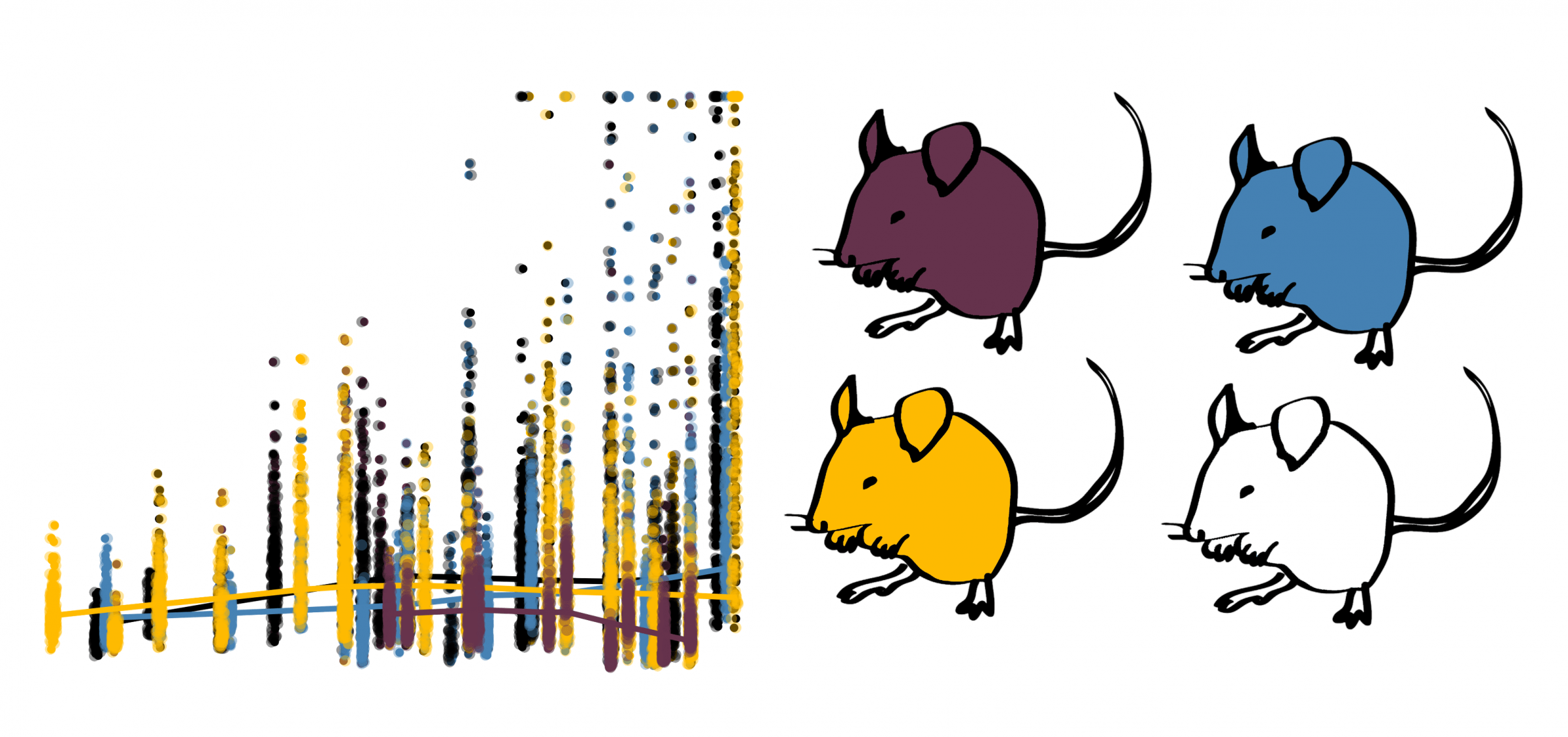
A multi-institute collaboration sought to map in high-resolution the earliest effects of the Huntington’s disease mutation in mice. The study, published Feb. 27, 2017, in Human Molecular Genetics, collected brain tissue weekly from more than 800 mice between 4 to 20 weeks of age in order to plot weekly changes in molecular phenotypes and gene expression that result from the disease mutation. A key feature of the study was that, while most of human disease research in mice relies on just one strain of mouse and therefore does not quantify variation that results from genetic background, this study included four different genetic strains of mice and found distinct differences in the rate of mutation-induced changes.
Huntington’s disease is caused by a single mutation in the huntingtin (HTT) gene. It was the first human genetic disease mutation to be characterized at the DNA level – before we had even sequenced the human genome. While the mutation is easy to understand, the disease characteristics are not. Part of what researchers have been trying to understand over the past few decades is the mechanism by which the disease mutation causes massive neurodegeneration in the brain (typically during middle age). 
Understanding key mechanisms in HD has been hindered by two things. One is that when studying human brain diseases in the human brain, researchers often must rely on post-mortem tissue that has reached the very last stages of disease. In late stage disease tissue, we observe thousands of genes that have changed expression and massive neurodegeneration. This makes it very difficult to understand the first effects. Another hindrance to understanding key mechanisms is the availability of data that tracks disease effects over time. Many recent studies in HD mouse models have attempted to check which changes have occurred at earlier time points and at multiple ages in mice. But this is the first study to track those changes weekly from such a young age. This allowed the researchers to capture which genes change expression first and at what age, and how the timing of those changes relates to other phenotypes such as mutant protein aggregation.
This study was a major collaborative effort across seven research groups including the Institute for Systems Biology, the McLaughlin Research Institute, the CHDI Foundation, and the Center for Human Genetic Research at Massachusetts General Hospital. The researchers hope that the publicly available data generated for the study will be used by other researchers in the field to further understand the early mechanisms of Huntington’s disease.
Journal: Human Molecular Genetics
Title: High resolution time-course mapping of early transcriptomic, molecular and cellular phenotypes in Huntington’s disease CAG knock-in mice across multiple genetic backgrounds
Authors: Seth A. Ament, Jocelynn R. Pearl, Andrea Grindeland, Jason St. Claire, John C. Earls, Marina Kovalenko, Tammy Gillis, Jayalakshmi Mysore, James F. Gusella, Jong-Min Lee, Seung Kwak , David Howland, Minyoung Lee, David Baxter, Kelsey Scherler, Kai Wang, Donald Geman, Jeffrey B. Carroll, Marcy E. MacDonald, George Carlson, Vanessa C. Wheeler, Nathan D. Price, and Leroy E. Hood
Link: academic.oup.com/hmg/article-abstract/doi/10.1093/hmg/ddx006/3045004/High-resolution-time-course-mapping-of-early


 isbscience.org/research/mapping-early-effects-huntingtons-disease-mutation-mice/