Closing a Critical Gap in Understanding Infectious Bacteria
How bacteria – including infectious ones– coalesce into assemblages called biofilms that help them resist antibiotics, is not well understood. Assistant Professor Anna Kuchina invented a technology (microSPLiT) allowing researchers to individually study hundreds of thousands of bacteria in a single experiment. The recipient of a promising investigators NIH grant (MIRA), her Lab focuses on closing the gap in our understanding of bacterial biofilms

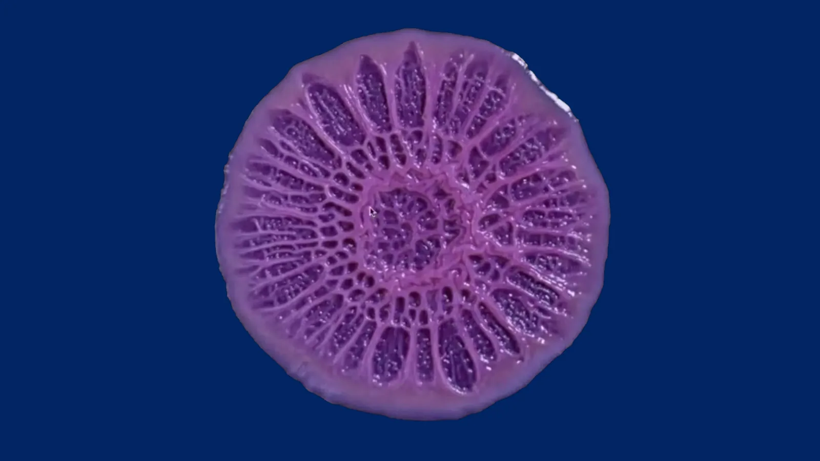
Still from a time-lapse video of growing Pseudomonas aeruginosa biofilm. Image credit: Scott Chimileski.
Although bacteria are commonly thought of as simple, single-celled creatures, many of them actually form complex and diverse communities. Known as biofilms, these structures are composed of multiple individual bacterial cells, sometimes from different bacterial species. Biofilms are the predominant natural state of bacteria and for infectious pathogens, they can be far more resistant to antibiotics than free-floating bacterial cells. ISB’s Anna Kuchina, PhD, is leading a project to better understand the biology of bacterial biofilms, using a novel technique she developed, known as microSPLiT (microbial Split-Pool Ligation Transcriptomics), to study the molecular details of the many individual bacterial cells that make up a biofilm community.
- Funded by National Institute of General Medical Sciences
- Led by Anna Kuchina, PhD
- Key collaborators:
- Nitin Baliga, MsC, PhD; ISB
Understanding how bacteria form drug-resistant biofilms
Kuchina developed the microSPLiT technique during her postdoctoral fellowship at the University of Washington, prior to joining ISB. At that point, no other methods existed that allowed researchers to characterize the expression of any gene across the genome in individual bacterial cells. The novel method enables researchers to see how all genes across the genome are switched on or off in a single bacterial cell across hundreds of thousands of individual cells in a single experiment. Based off a similar technique developed for mammalian cells, called SPLiT-seq, microSPLiT uses combinations of molecular barcodes to label individual cells so they can be distinguished from each other even if pooled together. Unlike techniques that separate and analyze one cell at a time using expensive instruments, the SPLiT methods can be performed with regular lab equipment and hence are relatively low-cost, enabling access to cutting-edge single-cell technologies for more research labs.
For Kuchina’s work, this single-cell approach is imperative to studying the differences among bacterial cells of the same species in a biofilm. Previous methods that involved capturing the average gene expression of a group of bacterial cells or biofilm would not allow the kind of detailed analysis needed to understand bacterial cell diversity within a community.
At ISB, Kuchina is leading a project to understand how the bacteria Pseudomonas aeruginosa forms biofilms together with another bacterial species, Staphylococcus aureus. Although these two bacteria are toxic to each other when free-floating in a test tube, they can actually form a cooperative biofilm in some environments, including in human lungs during chronic infections. These infections are especially tenacious in cystic fibrosis patients, who have weakened immune systems; biofilms composed of both bacterial species together may be more resistant to antibiotics than either bacterium alone. Better understanding how these bacteria interact in biofilms could point to new treatment avenues for these chronic co-infections, as well as shed new light on the general biology of biofilm communities.
Kuchina and her laboratory team have established a method to grow and study biofilms in the lab using a special medium that mimics the environment of an immune-compromised lung. They are now studying Pseudomonas’ cellular diversity in biofilms alone and will ask how gene expression and bacterial populations in the species change when the second bacterial species, Staphylococcus, is added to the biofilms.
In addition to studying gene expression in individual cells with microSPLiT, Kuchina and her team are also using microscopy imaging to understand the behavior of different populations of bacterial cells within biofilms. Because microSPLiT involves dissociating the biofilm into individual cells, the method can’t reveal spatial information about different kinds of cells within the community. To address this question, the researchers will use microSPLiT to identify specific genes switched on in different populations of cells and then build fluorescent reporters using those genes to light up different kinds of cells in the biofilms. This will allow them to follow the bacterial populations over time, gaining a sense of the dynamics of their growth, as well as to see where the populations exist in relation to each other within the biofilm.
Citations
- Gaisser KD, Skloss SN, Brettner LM, Paleologu L, Roco CM, Rosenberg AB, Hirano M, DePaolo RW, Seelig G, Kuchina A. High-throughput single-cell transcriptomics of bacteria using combinatorial barcoding. Nat Protoc. 2024. doi: 10.1038/s41596-024-01007-w.
- Kuchina A, Brettner LM, Paleologu L, Roco CM, Rosenberg AB, Carignano A, Kibler R, Hirano M, DePaolo RW, Seelig G. Microbial single-cell RNA sequencing by split-pool barcoding. Science. 2021. doi: 10.1126/science.aba5257.
Technologies Developed
microSPLiT (microbial Split-Pool Ligation Transcriptomics)

Contact Dr. Anna Kuchina
Assistant Professor
ISB